- New BESA workshops scheduled!
Find more information here!
Overview
BESA Research 7.1
Choose the best signal processing for your EEG and MEG data
BESA is the most widely used software for source analysis and dipole localization in EEG and MEG research. BESA Research has been developed on the basis of 30 years experience in human brain research by the team around Michael Scherg, University of Heidelberg, and Patrick Berg, University of Konstanz.
BESA Research is a highly versatile and user-friendly Windows® program with optimized tools and scripts to preprocess raw or averaged data for source analysis and connectivity analysis. All important aspects of source analysis and source imaging are displayed in one window for immediate selection of a wide range of tools. The same holds true for the source coherence / time-frequency module, and other analysis windows. BESA Research provides fast and easy hypothesis testing, a variety of source analysis algorithms including cortical imaging and volume imaging methods, integration with MRI and fMRI, age-appropriate template head models (FEM) as well as the possibility to import individual head models (BEM and / or FEM) generated by BESA MRI.
New features in BESA Research 7.1 are described here.
Connectivity analysis, a unique feature for viewing brain activity
BESA Research transforms surface signals into brain activity using source montages derived from multiple source models or beamformer imaging. This allows displaying ongoing EEG/MEG, single epochs, and averages with much higher spatial resolution.
The BESA Connectivity solution and the Source Coherence Module provide an extremely fast and user-friendly implementation of time-frequency analysis based on wavelets and / or complex demodulation. Users can create event-related time-frequency displays of power, amplitude, or event-related (de)synchronization and several connectivity measures for the current montage using brain sources or surface channels. Induced and evoked activities can be separated. Source connectivity analysis reveals the functional connectivity between brain regions by reducing the volume conduction effects seen in surface coherence. Users can select a time-frequency window for (bilateral) beamformer and dynamic imaging of coherent sources (Gross et al., PNAS 2001).
More than just dipole source localization
BESA Research covers the whole range of signal processing and analysis from the acquired raw data to dynamic source images:
MATLAB interface

Certification
BESA is CE Certified
![]()
Here you can download the Conformity declaration for BESA Research 7.1: PDF (71 KB)
EEG Review & Mapping
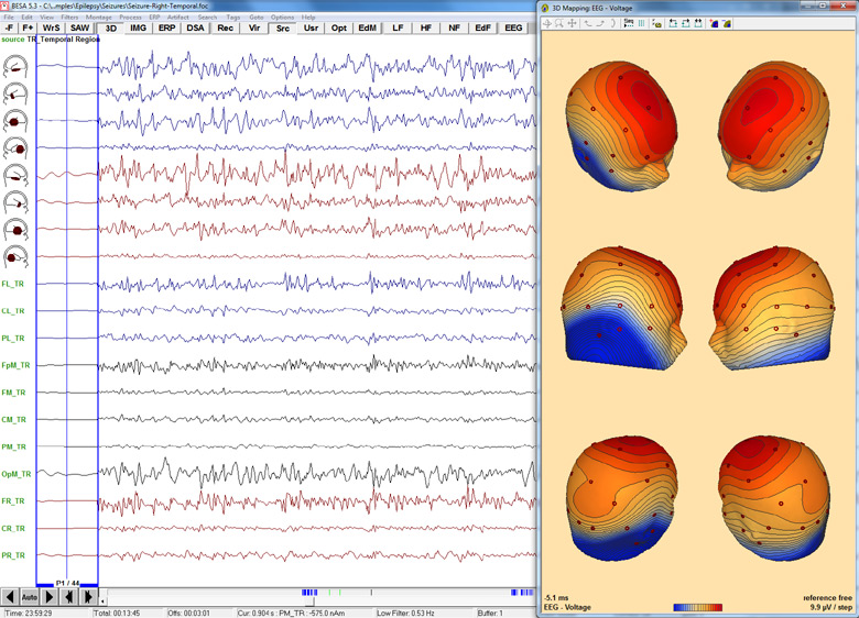
Onset of epileptic seizure with 3D whole-head maps, source montages, and average over onset cycles.
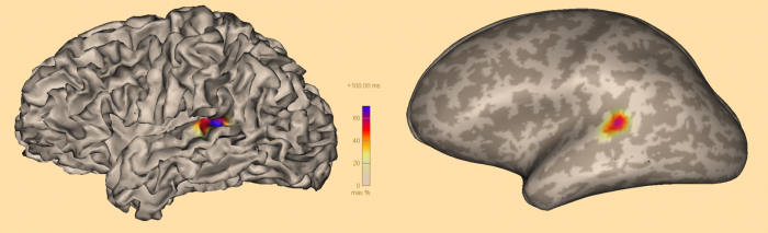
Source Analysis
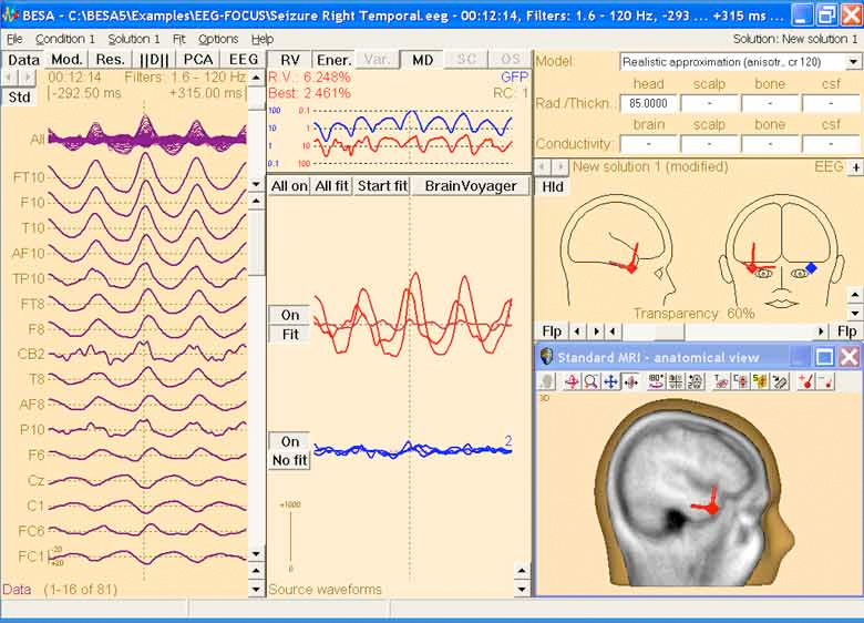
Source analysis at seizure onset using a FEM standard realistic head model and regional sources for the right and left temporal lobes.