- BESA Research 7.1 May 2025 released
It is a maintenance release with important bugfixes and a few vital improvements. Download it now here!
Features in BESA Research 7.0
Features in BESA Research 7.0
BESA Research 7.0 sets new standards in several aspects of neurophysiological data analysis: Welcome to the most streamlined and comprehensive analysis package available for state-of-the-art research using human EEG and MEG data! Particular highlights of the new feature list include:
Connectivity analysis

• Stand-alone module with 64-bit architecture and modern workflow design
• Use wavelets and / or complex demodulation
• Analyze connectivity in source space or sensor space
• Latest connectivity methods including Granger Causality, Partial Directed Coherence, …
• Visualize data in clear 2D and 3D result plots and create publication images or videos
• Seamless integration with BESA Research
• Export results for further analysis in e.g. BESA Statistics
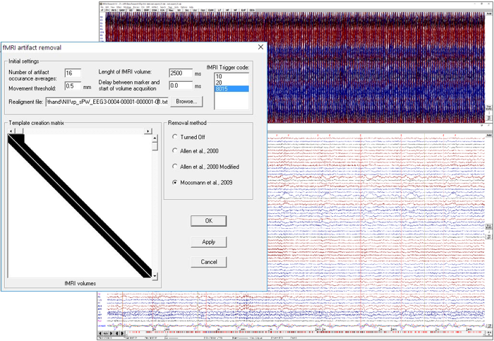
Simultaneous EEG-fMRI imaging

• Correct your fMRI artifacts directly in the BESA Research review window
• Choose between three proven methods for correction with few mouse clicks
• Read your fMRI data directly into BESA Research
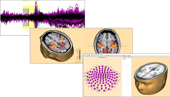
• Seed sources from fMRI and directly see activation patterns on millisecond scale
Beamforming

• Time-domain beamformer using one of several state-of-the-art methods
• Virtual sensor mode to reconstruct time courses
• Source montages using the beamformer spatial filter can be applied to raw data
• Compute beamformer image for any time point in interval of interest
• Conveniently select beamformer intervals graphically in ERP module
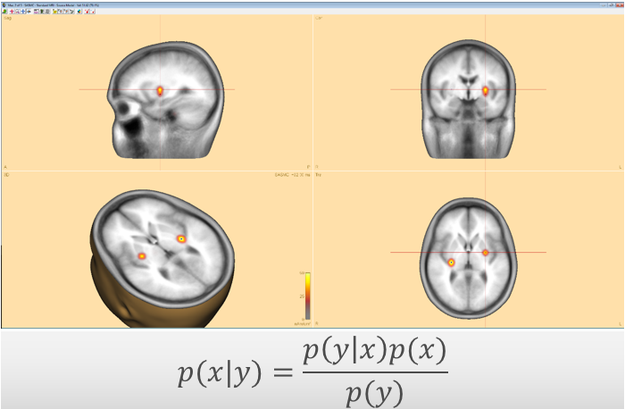
Bayesian source imaging

• Sequential Semi-Analytic Monte-Carlo Estimation (SESAME) of sources
• Automatically find most likely number of sources
• Compute map of likelihood for source positions
• Choose between most likely solutions for different numbers of sources
• Virtually no user input required
• Uses Markov-Chain Monte-Carlo method for efficient computation of probability distribution
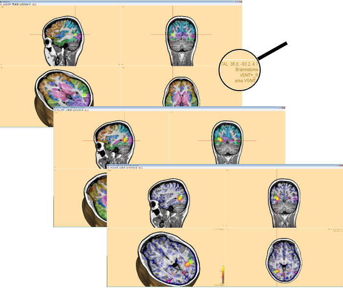
Brain atlases

• Check the anatomical or functional brain regions of your source imaging or dipole fitting results
• Seed sources from known locations in the brain atlas
• Choose between several state-of-the-art atlases including AAL, Brainnetome, Brodmann, …
• Visualize source imaging results together with atlas images
• Innovative contour display mode to facilitate brain image review

Recent Comments