This site presents the products of BESA GmbH, the leading innovators in digital EEG and MEG software for research and clinical applications.
- New workshops scheduled
New in-classroom workshops scheduled - more information here
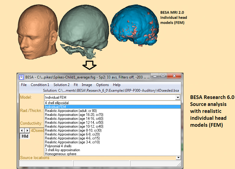
Individual FEM head model
Create individual FEM head models with BESA MRI
Individual, realistic volume conductor models improve EEG and MEG source analysis accuracy. BESA MRI in combination with BESA Research offer a complete pipeline for creating and using individual, realistically shaped head models for EEG and MEG source analysis. Minimal user interaction is required, and computation speed is highly optimized.
FEM head model computation in BESA MRI and use in BESA Research
- Segmentation of skin, skull, CSF and brain based on MRI grey values and tissue probability atlas
- Geometry-adapted hexahedral FE-mesh generation
- Forward computation for individual source space and given electrode / sensor configuration
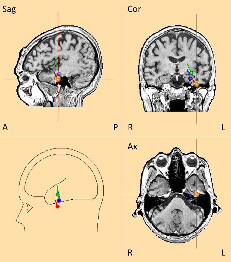
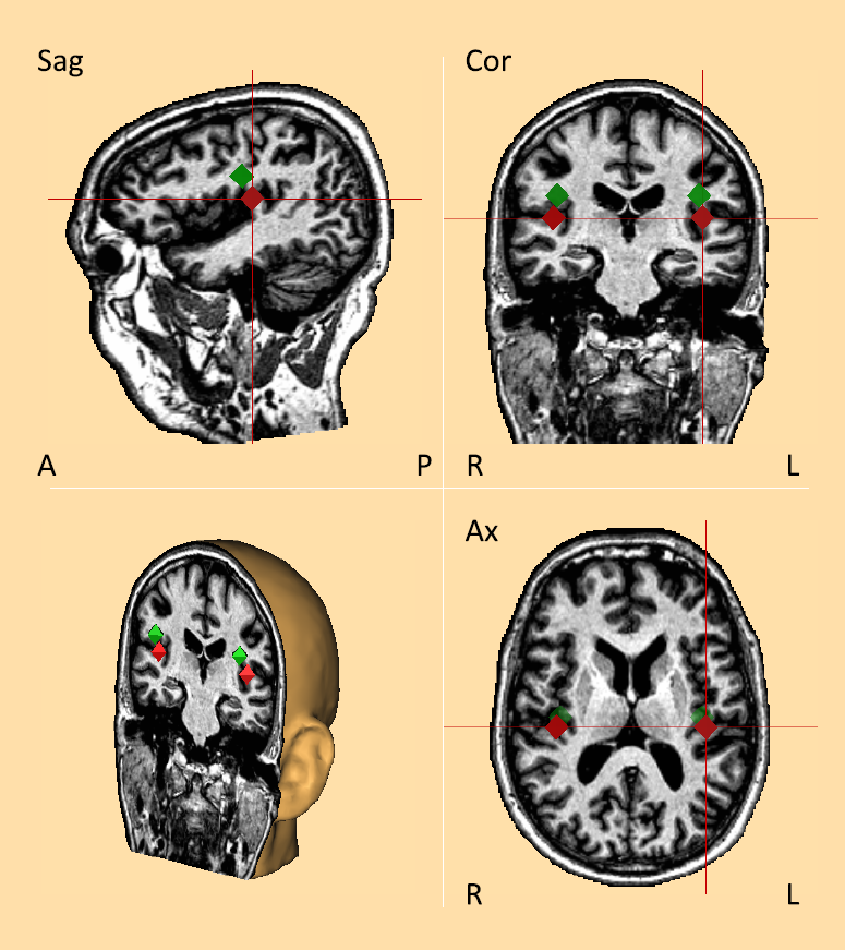
Improved source localization of epileptic spike and auditory activity with individual FEM
 |
 |
|---|---|
| Basal temporal lobe epilepsy – dipole sources reconstructed from the interictal EEG. Green: 4-shell ellipsoidal model; blue: standard FEM model; red: individual FEM model | Auditory N1 – Heschl’s Gyrus activity. Green: 4-shell ellipsoidal model; red: individual FEM model |
Download poster on BESA FEM pipeline
Individual FEM pipeline for EEG source analysis requiring minimal user intervention – OHBM meeting 2014
Recent Comments