Integration with MRI and fMRI
Integration with MRI and fMRI
For easy source analysis using individual MRI data, BESA Research provides an interactive interface to BESA MRI. EEG / MEG data can be coregistered with individual MRI data using individually digitized electrode positions or standard electrodes.
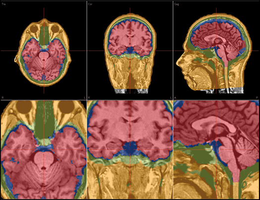
Coregistration and FEM model generation in BESA MRI

BESA MRI
- Integrated workflow for all user-interactions
- Automatic preprocessing with inhomogeneity correction
- Automatic reconstruction of scalp and cortex
- Automatic generation of individual 4-layer FEM model (scalp, skull, CSF and brain)
- Co-registration using digitized electrodes / headshape points or 10-10 / 10-20 standard electrodes
- Generation of FEM leadfields for individual head models co-registered with EEG electrodes and / or head surface points
- Overlay of source reconstructions on the individual MRI in BESA Research
- Separate license required for BESA MRI program
BESA Research also provides an interactive link to Rainer Goebel’s BrainVoyager™ (BV) program. The bidirectional connection of the two programs allows for source seeding from fMRI clusters with one mouse click.
BrainVoyager™
- Direct and easy interactive user interface of BESA Research with BV
- Analysis of individual MRI and fMRI data in BV
- Visualization and processing of individual MRI and fMRI
- Automated rendering of scalp and cortical surfaces
- Expansion and flattening of the cortical surface
- Minimum norm current image based on individual gray/white matter boundary
- Seeding of sources into BESA Research from anatomical 2D or 3D MR images or from fMRI BOLD clusters in BV via interactive link
- Overlapped display of fMRI and EEG / MEG sources in BV
- Separate license required for the BrainVoyager™ program

Recent Comments